A List of Pregnancy Scans and Their Benefits
Pregnancy Scans
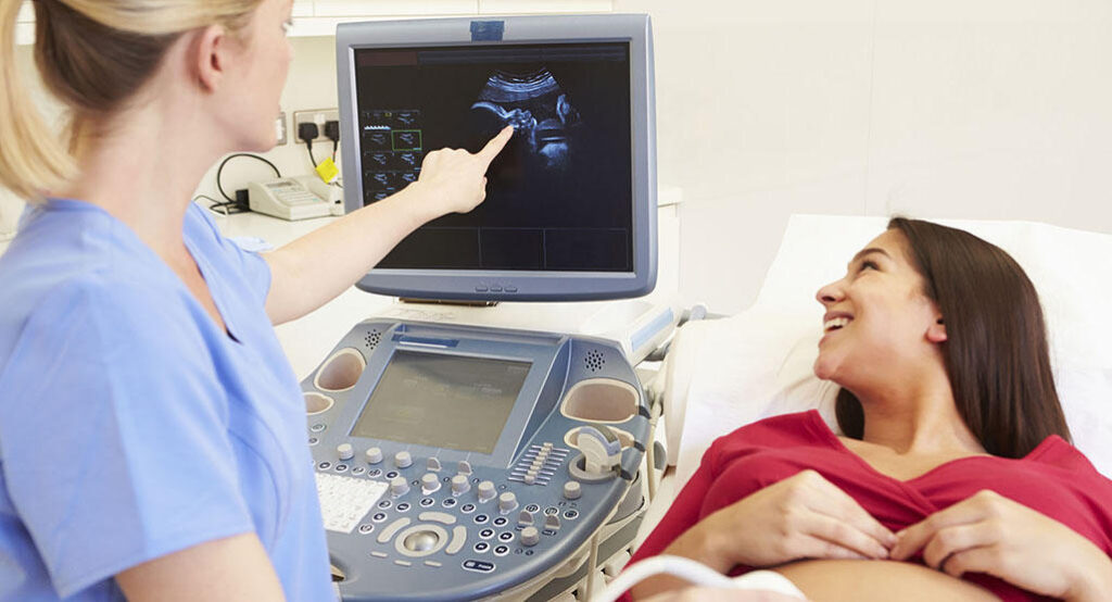
Expectant mothers need to be regularly scanned throughout their pregnancy period. The first scan is done during the first trimester to confirm the due date. The next scan is a growth scan done between the 18th and 22nd weeks to see if the foetal anatomy is normal. In all these cases, four ultrasound scans are performed. Apart from these, several other scans are also coming to the fore as technology improves.
Here is a list of the most important pregnancy scans and their uses.
Viability Scan
It is an early scan performed to confirm pregnancy, record the heartbeat and give the estimated due date. A viability scan is carried out between the 8th and 14th weeks of pregnancy. The sonographer will calculate the baby’s age by measuring the baby. The scan helps check if it is growing in the right position and verifies multiple foetuses.
Nuchal Translucency Scan
This pregnancy scan is carried out between the 10th and 14th weeks of pregnancy to examine the baby’s development and growth. Nuchal translucency is determined by measuring the fluid at the back of the neck of the baby. This helps assess the risk of different conditions like Patau’s, Edward’s and Down’s Syndrome. Blood tests are often needed for further analysis.
Reassurance Scan
It is an optional scan suggested for anxious parents who have complications like a high-risk pregnancy. The scan is performed if the expecting mother has health problems like bleeding.
Anomaly Scan
It is done around week 20 to measure the baby and examine its limb structures, bowel, spine, heart and brain for any abnormalities. The scan also helps determine the performance of the umbilical cord, the amount of amniotic fluid and the functionality of the placenta.
Cervical Scan
It is an extra transvaginal scan done in a pregnancy hospital to help measure the mother’s cervical length. The scan is performed on patients who carry the risk of giving a preterm birth.
3D Scan
It is performed between and 26th and 30th weeks to show the baby’s real-life image. Through the scan, the sonographer can find out the exact depth, height and width of the baby. The baby’s external body images are received in the scan. They show its facial features.
4D Scan
It offers a video rather than still images and captures the highlights and shadows. The baby’s movements and face are properly displayed through it.
No risks are associated with these scans. Ultrasound scans use sound waves to create an image of the baby in the mother’s uterus. Since they are non-invasive, they are painless and involve no side effects for the mother and the baby. Scans are an important part of any pregnancy to ascertain foetal well-being and health and the estimated due date. Scans help terminate the possibility of any health condition of the foetus. If the pregnancy comes without any risks, a regular scan is scheduled at certain intervals. Make sure to visit a reputed pregnancy hospital near you where you can have all the important ultrasound scans.